


The eukaryotic cell,

Introduction[1]
All living organisms are composed of one or more cell types. In multicellular organisms, different cell types are united in tissues and organs, which also include extracellular matrix (ECM) and interstitial fluid[2]. A cell can be defined as: 'the smallest, organised living unit within an organism, which, thanks to a complex metabolism, can exist more or less independently in a physiological environment and is capable of movement, growth and division by mitosis'. At the level of organisation, the cell is situated between the level of the molecules and that of the tissues, in a range that can be defined as follows in the classification of biomedical disciplines: molecules, cells, tissues, organs, individuals and society. Different cell types can act together as building blocks of a tissue. More than two hundred different cell types are counted in the human body. Cells reach ages ranging from a few days to as long as an organism lives. They can be replaced, and are also constantly replacing their organelles and building blocks, the proteins, lipids and carbohydrates, and combinations of these molecules. The synthesis[2] of proteins and enzymes needed for this is determined by the information derived from the genome.
All cells originated from a primitive primal cell through mutation and selection during evolution. Single-celled organisms, such as bacteria and protozoa[2], have adapted to the most diverse conditions and form more than half of the biomass on earth. In single cell organisms, all functions must be performed by a single cell. In a multicellular organism, cells can differentiate themselves by expressing different parts of their genome.
The cell types have a different structure and function, and can maintain a complementary collaboration with their different functions. As a result, multicellular organisms can perform more complex tasks than single-cell organisms, giving them an evolutionary advantage. The development of this complexity is accelerated over and over again with the development of an embryo from the fertilized egg.
An important step in evolution is the development of eukaryotic cells, in which the DNA[2] is separated from the cytoplasm and stored in a nucleus surrounded by a core membrane. For an overview of the main differences between prokaryotic and eukaryotic cells, see Table 1-1 and Figure 1-2.
Table 1-1 Significant differences between eukaryotic and prokaryotic cells.
Prokaryotic cell (e.g. bacteria, blue-green algae). | |
Unicellular or multicellular | Exclusively unicellular |
Diameter 5 - 100 µm | Diameter 0,5 - 10 µm |
Core (closed off from the rest of the cell by the nucleus envelope) contains genetic information from complexly organised chromosomes consisting of DNA + proteins | Genetic information in circular DNA located in the cell (nucleoid, genophore) |
RNA synthesis in core, protein synthesis in cytoplasm, nucleoli present in core | RNA and protein synthesized in the same compartment, no nucleoli |
Cytoplasm with cytoskeleton consisting of proteins | No cytoskeleton, organelles barely or not developed |
Organelles with specialized function in cytoplasm | |
Divide by mitosis or meiosis | Dividing by strapping |
Basically aerob[2] metabolism | Anaerobic[2] or aerobe metabolism |
Figure 1-2 Schematic illustration of a bacterium (A, prokaryotic cell) and a mammalian cell (B, eukaryotic cell).
A; The bacterium is about to divide, after the nucleoid (N) has doubled. The cell membrane is multiple and the cytoplasm contains no organelles.
B; The much larger eukaryotic cell, drawn on a different scale, shows many organelles or compartments secreted from the cytoplasm by a membrane. One recognizes a nucleus (N) with a nuclear body or nucleolus, the rough endoplasmic reticulum (R), the Golgi device (G) and mitochondria (M). The cell phagocytes[2] a bacterium (B), which will be digested after uptake into the lysosomes.

Eukaryotic cells are surrounded by a cell membrane and contain organelles, as separate, specialized compartments of the cell. The cell membrane is sometimes referred to as a plasma membrane or plasmalemma. Organelles are separated from the surrounding cytoplasm by a membrane and each has its own specific structure and function. Prokaryotic cells lack this organisation and are therefore less differentiated. Eukaryotic cells originated from prokaryotes, when the concentration of oxygen on earth began to increase. The size of an eukaryotic cell can vary greatly, from 3-5 µm to more than 100 µm. The cells of an elephant are not larger than the corresponding cells of a mouse, but the elephant has more. Cells with a similar function usually also have a very similar structure. Cells with an equal structure almost always have a similar function. When exceeding a certain 'critical mass', a cell will start to divide (mitosis). This critical mass is unequal for different cell types, and sometimes unequal for a cell in different phases of life. Cells can fuse and then form a syncytium, a multicellular cell. Multicellular cells can also arise after core division (karyokinesis), in which the division of the cytoplasm (cytokinesis) is omitted, resulting in a symplasma.
Parts of the cell[1]
In the nucleus of a cell, which is only missing in red blood cells and platelets, the genetic information is stored in intact but non-condensed chromosomes. The cell, as a whole, consists of the protoplasm, while cytoplasm is the part of the cell outside the nucleus. In the cytoplasm we find the following organelles, which are important compartments of the cell:
1. mitochondria, which are responsible for supplying energy;
2. endoplasmic reticulum, which is responsible for protein synthesis[2];
3. Golgi complex, which plays a role in the formation of secretion granules;
4. lysosomes, which take care of the intracellular digestion.
All organelles are surrounded by a membrane and contain specific enzymes that support their function.
The cytoplasm[1]
By dividing the protoplasm into separate compartments, as formed by the organelles, various metabolic processes can proceed in parallel (e.g. synthesis next to decomposition). The organelles and inclusions of the cytoplasm are embedded in the cytosol, the basic liquid substance of the cytoplasm. The inclusions form temporary constituents of the cytoplasm, and can consist of small accumulations of lipids, carbohydrates (glycogen) or pigment granules. In addition to the organelles and inclusions, there is also the cytoskeleton, including centriles, microfilaments, intermediate filaments and microtubules.
The cell membrane[1]
The plasma membrane or cell membrane is composed of phospholipids, cholesterol, proteins and glycoproteins. The plasma membrane acts as a selective barrier, regulating permeability and transport between the cytoplasm and the extracellular environment. The membrane can pass substances passively or transport (active) substances over the membrane using energy. The plasma membrane and the cell coating or glycocalix present on it have a function in recognizing and possibly attaching substances, particles and neighbouring or foreign cells. The plasma membrane and the cytoskeleton also play a role in the movement of cells.
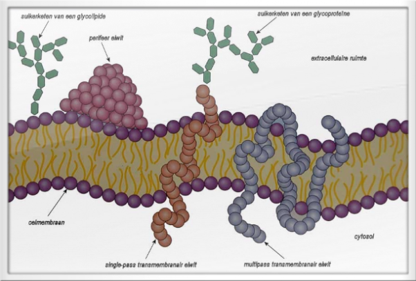
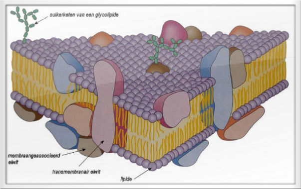
All membranes of the cell show a unit structure (unit membrane), mainly composed by a phospholipid double layer, which is visible in a transmission EMI image as two parallel lines with a distance between 5 and 10 nm (fig 1-3). In such a membrane, the hydrophobic tails of the phospholipids[2] lie towards each other, and the hydrophilic parts form the outer layer of the membrane. Membranes are more or less liquid, i.e. a large part of the molecules in the membrane is freely movable. One can measure this 'fluidity' with certain methods.

Mitochondria[1]
Mitochondria are visible in living cells with phase contrast microscopy. They occur in all eukaryotic cells and can sometimes take up to almost half of the cytoplasmic volume, but usually this is much less. The number of mitochondria per cell varies from cell to cell and depends on the energy requirement. Blood lymphocytes have a few dozen, but a parenchyma cell of the liver has 2-3000, while an egg cell has several hundreds of thousands. Mitochondria are  usually elongated, sometimes branching, and have dimensions from 0.5 to a few µm.
usually elongated, sometimes branching, and have dimensions from 0.5 to a few µm.Mitochondria move within the cell and can undergo significant changes in shape. Mitochondria can also split and merge. Mitochondria convert the chemical energy of metabolic substances into ATP[2]. From ATP, energy can easily be released anywhere in the cell for energy-consuming processes, such as osmotic, mechanical, electrical or chemical work, ion transport or signal transduction. Mitochondria can concentrate at a place in the cytoplasm where a lot of energy is consumed, such as in the apical cytoplasm of vibratory cells, the middle segment of spermatozoa or the basal cytoplasm of ion transport cells.
The structure of the mitochondrion is well expressed in an electron microscope (Figure 1-4). Mitochondria have an outer and an inner membrane, which is strongly enlarged and folded into so-called cristae. The cristae are usually leaf-shaped, although there are also tubular cristae and steroid-forming cells. The double membrane of the motochondrion divides the organelle into a number of different compartments and surfaces, which have different functions:
1. the outer membrane;
2. the intermembranous space, which is the compartment between the outer and inner diaphragm;
3. the intramembranous surface of the inner membrane containing the cristae;
4. the matrix.
The outer membrane contains proteins for the transport and conversion of substrates[2], and is rather permeable, so that the composition of the intermembranous space largely corresponds to that of the cytoplasm. The inner membrane is less permeable, three-quarters composed of proteins, including transport proteins and the enzyme complexes of the respiratory chain. Some of these enzymes can be made visible as spherical elementary bodies in an EM. Sometimes matrix granules can be seen, which on analysis appear to contain calcium and magnesium salts or belong to the mitochondrial ribosomes. In the mitochondria, energy-containing raw materials are broken down by the enzymes of the citric acid cycle (Krebs cycle). The energy released is then used for oxidative phosphorylation. ATP is the final product, while CO2 and water are released. The enzymes that produce ATP are located in the inner membrane, while the matrix contains enzymes for the citric acid cycle. In the brown adipose tissue, mitochondria can decouple the oxidative phosphorylation and the electron transport and the energy is released as heat. Cells with high metabolic activity (e.g. cardiac muscle cells or renal tubule cells) have many mitochondria with densely packed cristae, while cells with low metabolic activity have a small number of mitochondria with few, short cristae.
Mitochondria contain a small amount of circular, double-stranded DNA in their matrix, which is not complexed with histones[2]. This DNA, via tRNA and ribosomes, provides its own protein synthesis, which is independent of nuclear DNA. This synthesis is only sufficient for about 10% of the mitochondrial proteins; the rest of the proteins are encoded in the nuclear DNA and transported from the cytoplasm to the mitochondrium. During cell dividing, mitochondria are more or less equally distributed among the daughter cells, which then replenish the stock. Multiplication of mitochondria takes place through cleavage, growth and DNA duplication. The fact that mitochondria contain their own circular DNA and ribosomes is reminiscent of the situation in bacteria. This has led to the so-called endosymbiont-hypothesis, which states that mitochondria are descended from bacteria that are included in the cytoplasm of eukaryotes during evolution in symbiosis[2].

Figure 1-4 TEM capture of a mitochondrion in a rat pancreatic cell.
The double membrane and the cristae (C) of the mitochondria (M) are clearly visible. If the cristae are not oriented perpendicularly in the cut, dark shadows appear instead of clear membranes (bottom-right). The RER is highly developed in these cells due to the very high production of digestive enzymes.
The endoplasmic reticulum[1]
The endoplasmic reticulum is a network (reticulum) of membranes located in the cytoplasm of a cell. It consists of two tightly packed membranes between which cavities and channels are formed. It is separated from the rest of the cell by a membrane with the same structure as the cell membrane.

The raw endoplasmic reticulum (RER or ribosomal ER) houses the ribosomes and therefore plays an important role in protein formation (protein synthesis) in the cell. It also has a role in the transport of substances in the cell, particularly important for the collection of proteins to be transported to the Golgi apparatus. This is actually the main function of the endoplasmic reticulum. The rough endoplasmic reticulum owes its name to the rough appearance under the electron microscope, which is caused by the many ribosomes attached to the outer surface. Ribosomes, due to their nucleic acid content in a classically stained HE (Haematoxylin-Eosin) coupe, produce a basophilic staining reaction (pink).
All attached ribosomes are connected to the inside of the ER by a pore protein. When one of these ribosomes receives a mRNA strand, the translational peptide chain is introduced through the pore protein into the ER. Only then is the protein folded to its final form.
The smooth ER (English: smooth ER, SER) is a variant of ER that mainly serves to transport substances from the crude ER to the Golgi apparatus; it is therefore often located between areas of crude ER and the Golgi apparatus. Other functions of the smooth endoplasmic reticulum are the storage of calcium ions, the synthesis of lipids, phospholipids and the detoxification of drugs, alcohol and other toxins (especially in liver cells). The SER is therefore well developed in the adrenal cortex (for the production of steroid hormones), liver parenchyma and muscle cells (where they play a role in the contraction process, the so-called sarcoplasmic reticulum).
References:
[1] Junqueira L.C. en Carneiro J. (2004, tenth edition), ISBN 13: 9789035226715, Functionele histologie, Maarssen. Uitgeverij Elsevier. Chapter 3, 'De cel'.
[3] Prof. Dr. med. Ulrich Welsch (2006, auflage 2), ISBN: 9783437444302, Lehrbuch Histologie, München. Uitgeverij Elsevier GmbH, Urban & Fisher.