Nissle substance in motor neurons, cresyl violet acetate staining
Histochemistry: the 'Nissl staining'.
Purpose of the slide: To visualize RER and free ribosomes in the cell body of neurons.
Neurons[1]
Nerve cells or neurons, when mature, are non-dividing cells that provide communication within the nervous system, as well as with other tissues (including muscles). They involve the uptake, transmission and processing of stimuli, involving neurotransmitters and other information molecules and role.
Neurons consist of three parts.
1. The cell body or perikaryon: the metabolic center of the cell, centered on the nucleus, which is also sensitive to stimuli.
2. The dendrites: highly branched offshoots, of which there are usually a number per neuron. They usually capture stimuli and conduct them to the cell body.
3. The axon: a single, often very long offshoot, which usually conducts impulses to other cells (neurons, muscle cells or glandular cells).
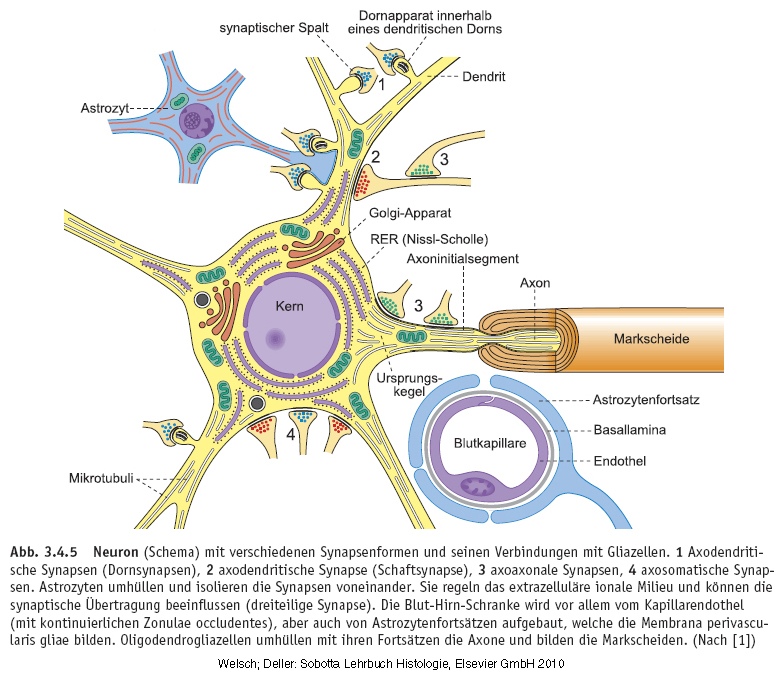

Perikaryon[1]
The perikaryon contains the nucleus of the nerve cell and the surrounding cytoplasm[2], without offshoots. This metabolic center can also receive impulses from other neurons on its surface.
The large nucleus has a prominent nucleolus[2] and finely divided chromatin. Also found are many free polyribosomes and a highly developed RER (rough endoplasmic reticulum[2]). All this indicates high protein synthesizing activity for structural and export proteins (neurotransmitters[2]). As early as the nineteenth century, RER and free polysomes[2], by staining with cresyl violet (Nissl staining), could be observed as basophilic elements in the cytoplasm: the Nissl substance. The amount of this substance varies according to the type of nerve cell and its activity. There is a lot of Nissl substance in large nerve cells, such as motor neurons[2].
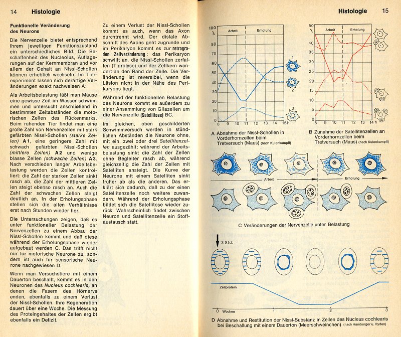
Changes in the perikaryon under varying conditions[3].
Prof. Dr. Med. Werner Kahle (Neurological Institute of the University of Frankfurt), describes in "dtv-Atlas der Anatomie"[3] the functional changes of a neuron that it can undergo under varying conditions such as, for example, performing labor or prolonged noise exposure.
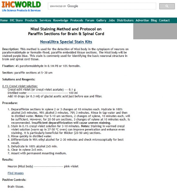
Staining of a slide with cresyl violet to demonstrate Nissl substance.
The staining is based on a protocol from IHCworld, see image to the right.
The author has adapted the protocol to his own slide but the principle remains unchanged.
However, it is very important to mention that the dye should be Cresyl violet acetate with CAS No: 10510-54-0.
The dye can be ordered for example from 'Merck'. See: https://www.sigmaaldrich.com/NL/en/search/10510-54-0?focus=products&page=1&perpage=30&sort=relevance&term=10510-54-0&type=product
Nissl stain | Times |
| Bring coupes in warm 35°C xyleen* | 10 min |
Bring coupes in warm 35°C xyleen* | 10 min |
Ethanol or isopropanol 100% | 5 min |
Ethanol or isopropanol 100% | 5 min |
| Ethanol 95% | 3 min |
Ethanol 85% | 3 min |
Ethanol 70% | 3 min |
| Ethanol 50% | 3 min |
Rinse in tap water | 5 min |
| Rinse AD | 5 min |
| Cresylviolet-acetaat 35°C | 30 min |
Rinse AD | 1 min |
Differentiate in Ethanol 95% (short) | ≈ 15 sec |
Isopropanol 100% I and II (no Ethanol)** | 2 x 4 min |
Xylol I and II | 2 x 4 min |
Malinol/Euparal/Depex*** |
* All paraffin should be well removed, ensures more uniform staining.
** Isopropanol stops the differentiation process better than Ethanol.
*** Depex is absolutely acid-free and sslides by the author are still unchanged after 10 years.
Cresylviolet-acetaat: |
- 0,1g Cresylviolet-acetaat dissolve in 100ml AD (warm till 60 graden Celcius); - After cooling, just before use, add 10 drops or 0.3 ml of Acetic Acid 100% and filter. |
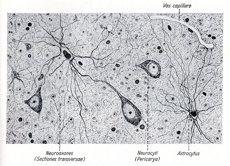
In the book "Atlas der normalen mikroskopischen Anatomie des Menschen"[4]...
...by Max Clara, Kurt Herschel and Helmut Ferner, various neurons are shown drawn. Here the intended Nissl bodies or "substantia tigroidea" are clearly visible.

Preparation details,
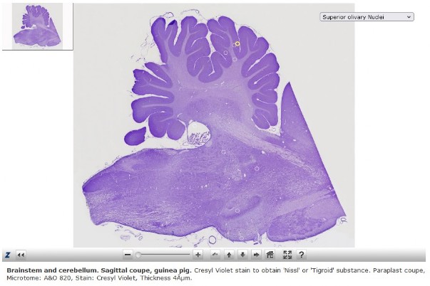
The brain stem and cerebellum were taken from a Guinea pig and fixed in formaldehyde 4%.
The block was embedded in paraplast plus and then cut on an A&O 820 rotary microtome. Thickness is 4µm.
Depex from the company Serva was chosen as mount resin and can be purchased there, among others: https://www.serva.de/enDE/ProductDet......DePeX_0_0.html
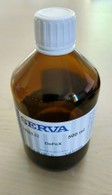 The resin is completely acid-free and easy to handle. After a few days of drying, excess resin can be easily removed with a Stanley knife.
To obtain a large-format image that is quite "zoomable" afterwards, the author took 171 shots with a Leitz plan Fluotar 16x objective. Used a Moticam 2300 camera on a Leitz Orthoplan microscope.
Click on the image to the right and zoom in on various structures of the brainstem and cerebellum.


References:
[1] Junqueira L.C. en Carneiro J. (2004, tiende druk), Functionele histologie, Maarssen. Uitgeverij Elsevier. Hoofdstuk 9, pag. 189-190, 'Zenuwweefsel', ISBN: 90 352 2671 2.
[2] Wikipedia, the free encyclopedie, https://en.wikipedia.org/wiki/Main_Page
[3] Prof. Dr. Med. Werner Kahle (1976), Taschenatlas der Anatomie für Studium und Praxis, dtv Verlag Munchen, Georg Thieme Verlag, Bd. 3., Nervensystem und Sinnesorgane, ISBN: 3 423 03019 4, ISBN: 3 13 492201 0.
[4] Prof. Dr. med. Max Clara (1974), Atlas der normalen mikroskopischen Anatomie des Menschen, Uitgeverij Urban&Schwarzenberg. pag. 357 en 373, 'Systema nervosum', ISBN: 3 541 06331 9.